bkoep Staff Lv 1
This is an extension of last week's protein design update, in which we discussed recent improvements in backbone quality and showcased a collection of player designs that were brought into the wet lab. Our analysis is ongoing, and some of those designs may still yield results. But a few exceptional designs are already showing promise, and we thought those results warranted a separate, more focused analysis here.
Folded Proteins
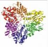
Below are four proteins designed by Foldit players, then expressed and purified in the Baker lab (more here). Experimental data from circular dichroism (CD) spectroscopy suggest that these proteins are stable and well-folded (figures explained in the key below).
Note that our testing is not yet complete—we still do not know whether these proteins are folding into their intended conformation or some other, alternative structure. For that we will need atomic-resolution data from x-ray crystallography or other methods.
 Susume (Anthropic Dreams) — Puzzle 1248
 Waya, Galaxie, Susume (Anthropic Dreams) — Puzzle 1297
 fiendish_ghoul — Puzzle 1299
 fiendish_ghoul — Puzzle 1299
Key:
(A) Cartoon diagram of each Foldit player-designed protein. All of these designs feature α-helices packed against a single β-sheet, but no two designs share the same fold.
(B) Rosetta@home folding predictions (described here). Rosetta@home was able to successfully predict the structure of each design based on its amino acid sequence. The "funneled" cloud of red points reaching toward the lower-left corner of each plot indicates that Rosetta is able to reconstruct the intended fold from sequence information alone, and that the intended fold is furthermore predicted to be the most stable.
(C) The circular dichroism (CD) spectrum of purified protein shows that each protein contains significant secondary structure. This characteristic CD signature—with a broad, flat trough between 208 and 222 nm—suggests that both α-helices and β-sheets are present at 25°C (blue trace). We see that most of this structure is retained at 95°C (red trace), and that lost structure can be recovered upon cooling back to 25°C (green trace).
(D) Each protein is fairly thermostable, retaining a strong CD signal at 220 nm as it is heated from 25°C to 95°C.
(E) These proteins are unfolded by titration of concentrated guanidinium chloride (a chaotropic agent). The steep, sigmoidal transition from the folded to the unfolded state suggests that each of these proteins folds via a cooperative, two-state mechanism.
Crystallization Trials
The next step is to try to crystallize these proteins. Under very specific conditions, a concentrated sample of purified protein will self-organize into a highly-ordered crystal lattice. Protein crystals are useful to us because they comprise a large number (think trillions) of identical protein molecules all locked into the same orientation. If we aim a focused beam of x-rays at a protein crystal, electrons in the ordered crystal lattice will diffract the x-rays to produce an ordered diffraction pattern. From this diffraction pattern we can infer the distribution of ordered electrons in the crystal at extremely high resolution, in the form of an electron density map, thus revealing the atomic structure of the crystallized protein.
Unfortunately, protein crystallization is a delicate process, and is very sensitive to subtle change in conditions. Different proteins require wildly different conditions for crystallization, and we have no way to predict which conditions will allow a particular protein to crystallize. Protein concentration, buffer, pH, salts, ligands, precipitants, temperature, and time can all be critical factors for crystal growth. Typically, a crystallographer will set up high-throughput crystal screens, incubating concentrated protein in large arrays with hundreds of different conditions, and monitor them over periods of weeks or months.
Ultimately, protein crystallization is a lottery. Many proteins are never successfully crystallized. But, with a little luck, we'll be able to grow crystals of some of these proteins, collect x-ray diffraction data, and determine their full structure.